Overarching Methodology and Conceptual Framework of the Research Program
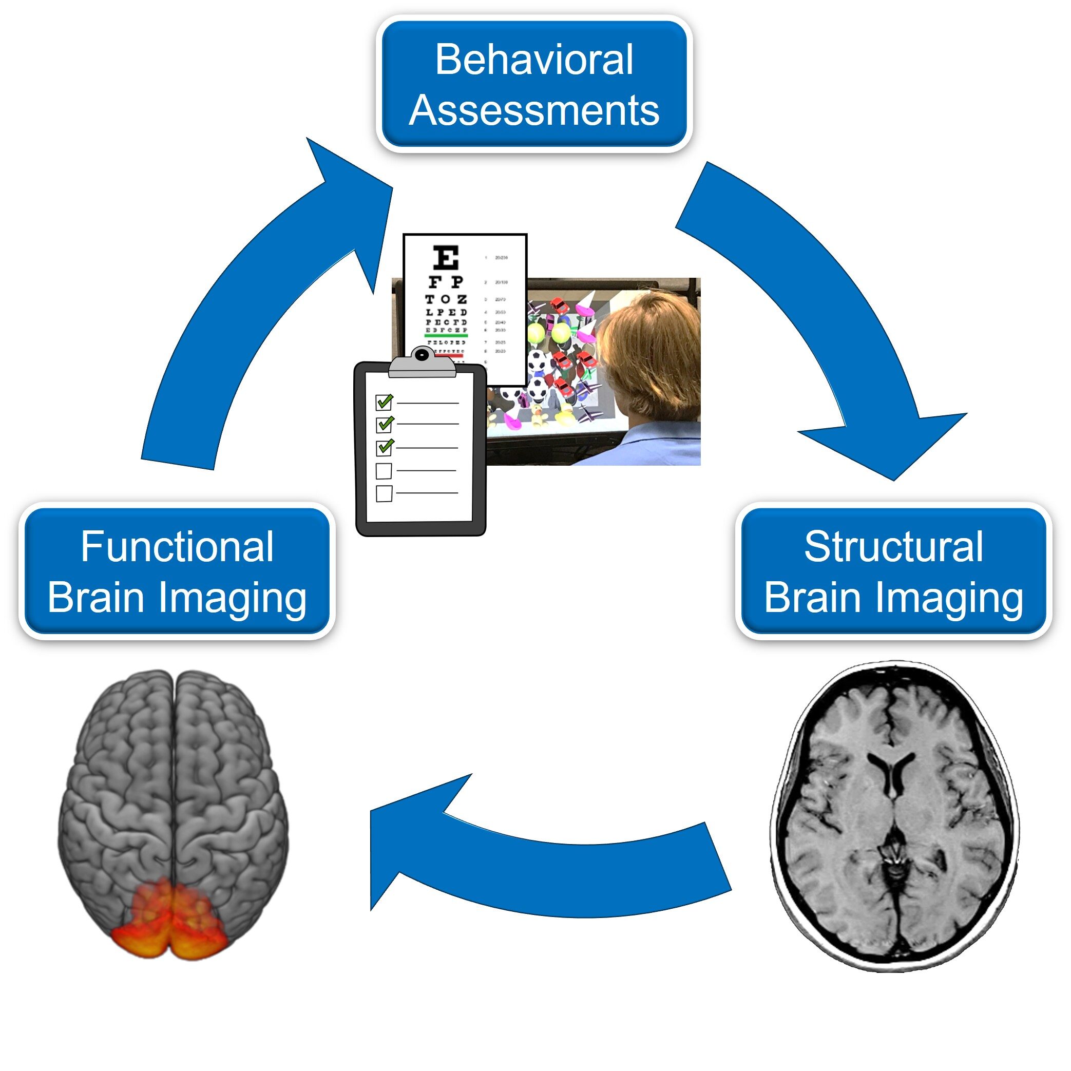
Our lab combines clinical behavioral assessments (e.g. visual function, functional questionnaires, and novel visual testing methods combining virtual reality and eye tracking), structural brain imaging (MRI and diffusion-based imaging), and functional brain imaging (fMRI, EEG, and resting state functional connectivity). Using a multidisciplinary approach, and investigating the association between various sources of data, we hope to have a better understanding of how visual behavior, brain structure, and brain function are interrelated.
Techniques Used
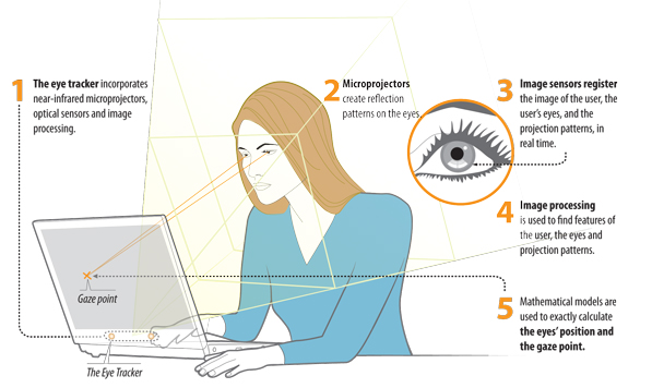
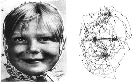
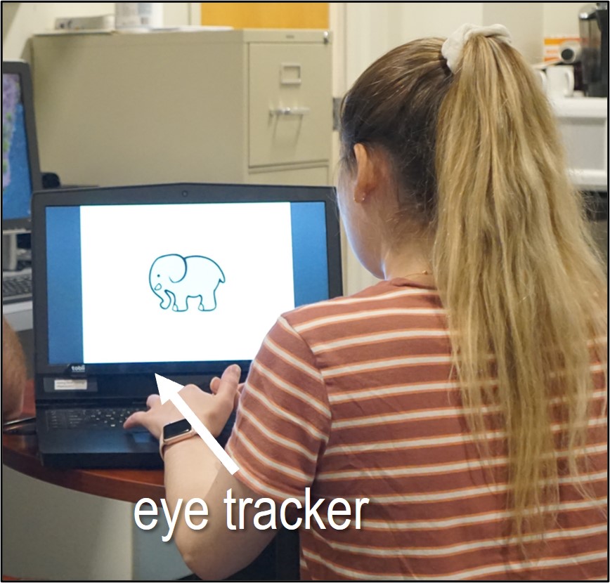
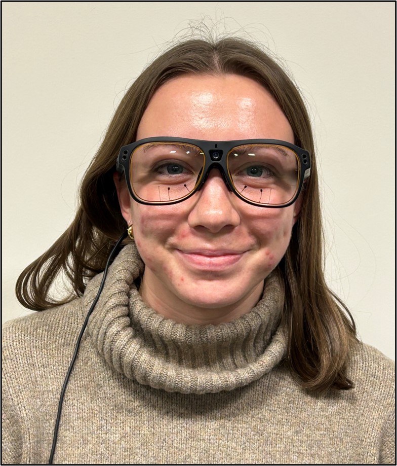
Eye tracking
Behavioral Testing and Cross-modal Sensory Interactions
Brain Imaging
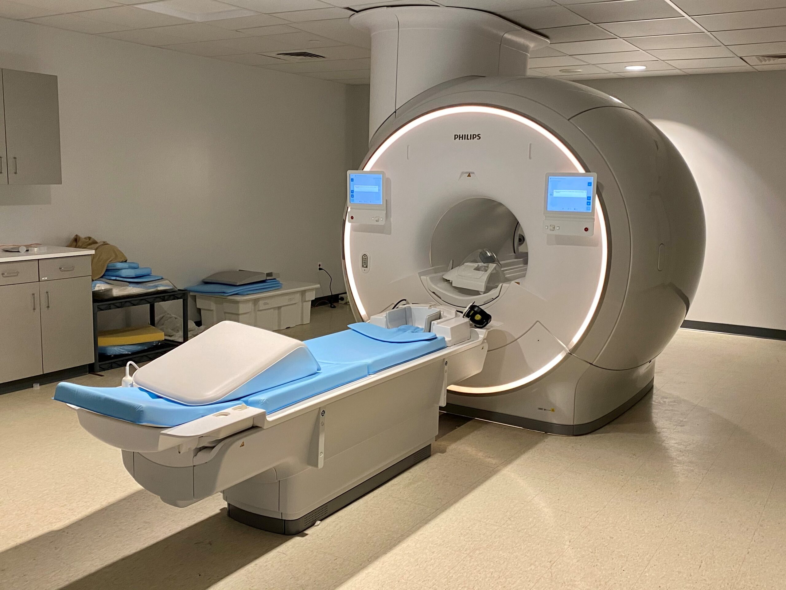
Structural neuroimaging allows for the visualization of the structure of the brain by highlighting the contrast between different tissues such grey matter, white matter, and cerebrospinal fluid.
Functional neuroimaging measures brain activity (directly or indirectly) associated with performance on a particular mental/behavioral task.
Current Projects
Visual Abilities and Neuroplasticity Associated With Cerebral Visual Impairment (CVI)
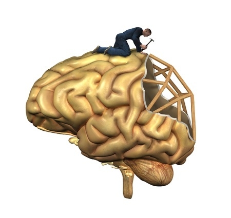
Cerebral (or cortical) visual impairment (CVI) is a brain-based visual and neurodevelopmental disorder and is the leading cause of visual impairment in children. It is characterized by visual dysfunction primarily associated with damage to central brain structures as opposed to the eyes. Despite this very important public health concern, the underlying neurophysiological basis of this condition remains poorly understood, and more research is needed to fully understand how the developing brain reorganizes itself in response to early damage and maldevelopment.
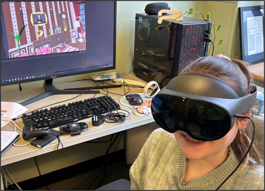
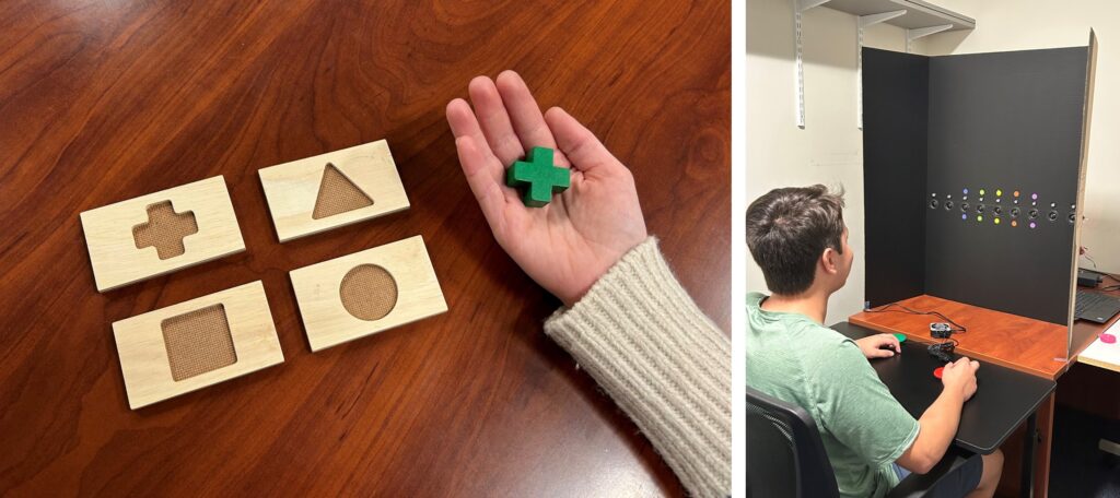
The goal of this investigation is to establish a conceptual framework relating visual and sensory functions with structural and functional reorganization in the brain. In this effort, we use high resolution structural imaging to reconstruct white matter pathways of the brain using diffusion-based imaging (specifically, High Angular Resolution Imaging, or HARDI). We carry out assessments integrating behavioral psychophysics, virtual reality (VR), and eye/gesture tracking technology. These novel techniques allow us to characterize visual performance beyond what is typically done in a standard clinical eye exam.
We share the results collected with our participants as well as their families, teachers, and health care providers to help further characterize the functional visual abilities of individuals with CVI.
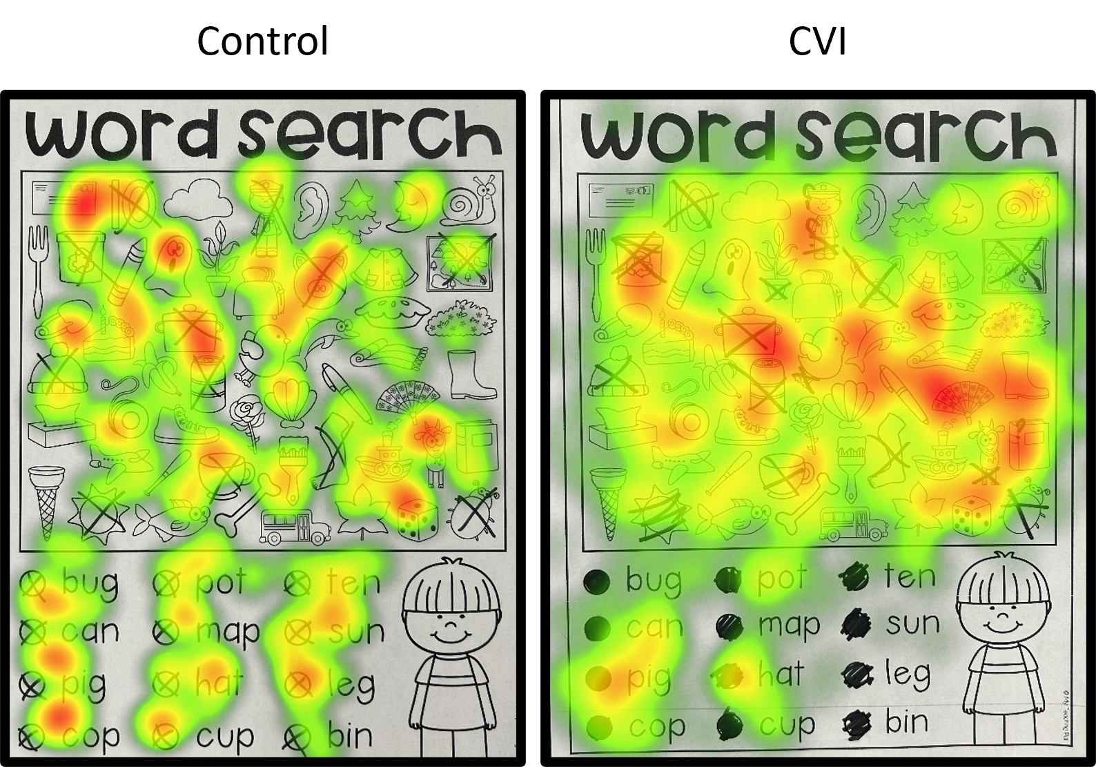
Gaze behavior (“heat maps”) recorded with wearable eye tracking showing the location of fixations during a word and picture search task. Note the overall pattern and extensive distribution of fixation points in the CVI (right) compared to control (left) participant. The more extensive gaze behavior may help explain the greater fatigue reported by individuals with CVI while carrying out class work and everyday tasks.
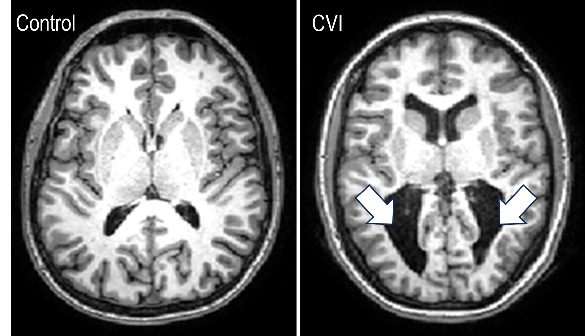
Brain structural changes revealed by MRI. Note the enlarged ventricles and surrounding structural changes (shown by the arrows) in a CVI (associated preterm birth and periventricular leukomalacia; right) compared to a control with neurotypical development (left).
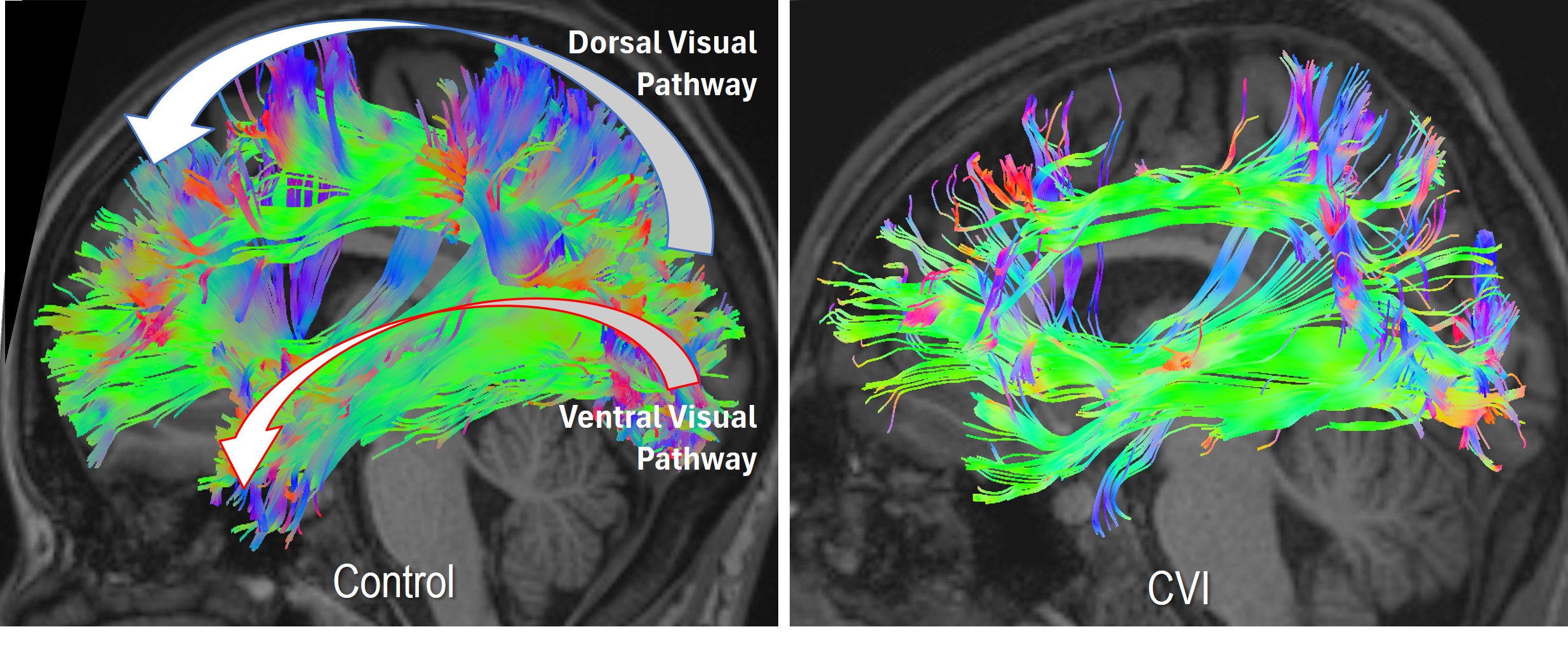
White matter reconstruction of the dorsal and ventral visual pathways using diffusion-based imaging (HARDI). Note how these pathways are not as well developed (particularly the dorsal pathway) in the CVI subject (right) compared to a control (left).
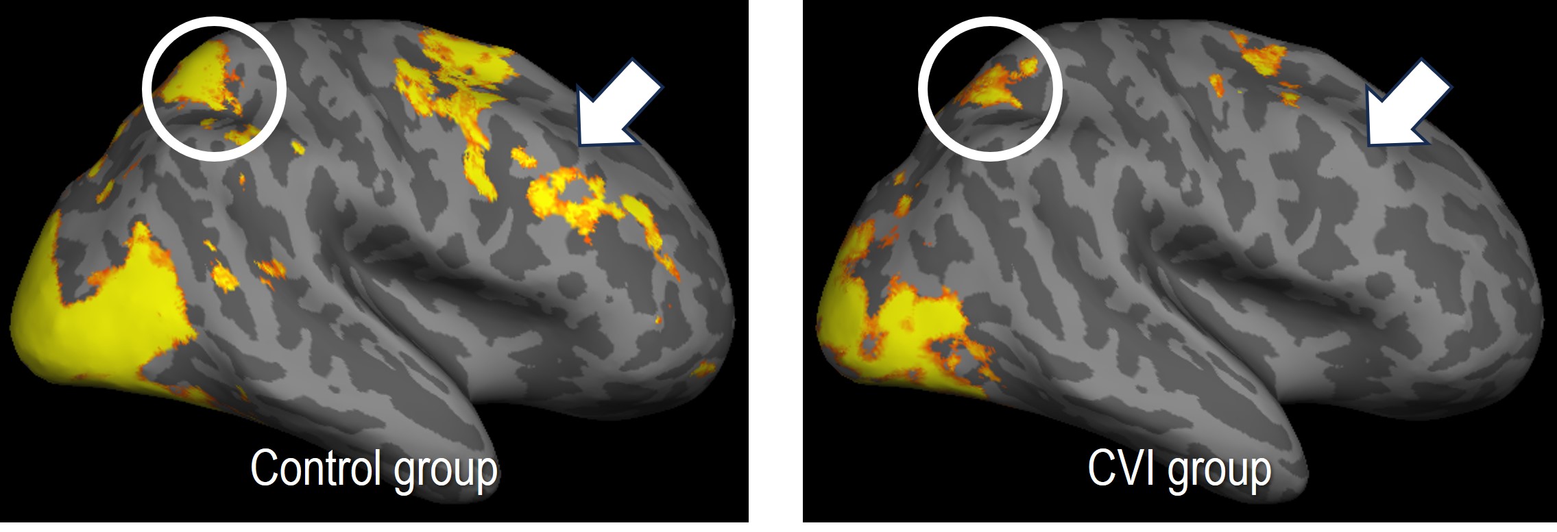
Activation patterns revealed by functional magnetic resonance imaging (fMRI) in response to a complex visual search task (only right hemisphere of the brain is shown). In controls (left), note the pattern of robust activation within the occipital visual cortex, parietal areas of the dorsal visual pathway (circle), and frontal cortical areas (arrow). Activation is comparatively less robust in the group with CVI (right).
